Diseases of the skin are unpleasant, and sometimeseven a frightening phenomenon. They deliver a lot of physical and psychological discomfort. Practically every person has some or other skin problems. However, the late removal of some of them can lead to serious complications. Intradermal nevus is one such problem.
general description

This pathology is oftena benign neoplasm on the skin, which mostly rises above its surface. In some cases, it can acquire tuberculate form, and sometimes remains small, differing in color stain.
Intradermal nevus has a painless surface and a soft structure. The color of the new growth can be different: pink, red, brown and even black.
An intradermal nevus can be located on theneck, scalp, face. Very rarely, education occurs on the body or limbs. It should be borne in mind that if the tumor is benign, then it practically does not cause painful sensations. In malignant form, melanoma, she is reborn in only 20% of cases.
If there is some discomfort in the area of skin lesions, or the intradermal nevus begins to grow rapidly, you should immediately consult a doctor.
Causes of appearance

So far, they have not been clarified for certain.But in any case, this pathology is a consequence of the abnormal functionality of the skin. There are factors that can trigger the development of the disease:
- Allergic skin diseases.
- Intrauterine disorders of fetal development.
- Heredity.
- Hormonal disorders, such as altering the body's work during pregnancy or menopause in women.
- Dermatological infectious pathologies.
Intradermal papillomatous nevus can also develop due to a strong toxic poisoning of the body, and the substance due to which it appeared does not matter.
Varieties of pathology

There are a lot of them:
- Galonevus.
- Blue. It has a relatively small size and a special blue color.
- Frontier. This formation is characterized by the fact that it rises above the skin surface only partially.
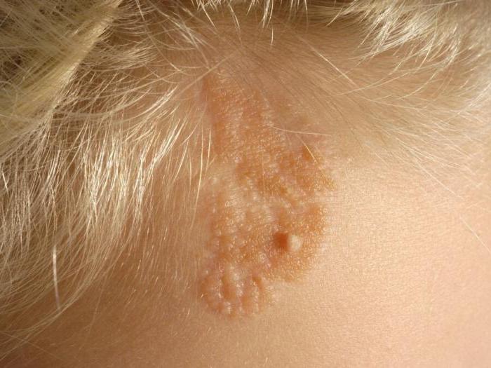
- Внутридермальный папилломатозный невус.Its size can exceed 1.5 cm, and over time it can grow even more. Most often it is a brown tubercle, similar to a wart. Inside the tumor, hard black hairs can be seen. With careful handling of this new growth, it rarely degenerates into a malignant one.
- Non-celled. It often appears on the face, causing serious physical discomfort. Its shape is usually convex. Because of its location, the tumor must be removed.
- Intradermal melanocytic nevus.It has a rich color, a correct, clear shape. It can be found on the chest and even on the genitals. The size of the lesion rarely exceeds 0.5 cm. It may be flush with the skin or slightly elevate above it.
Some of these formations can degenerate into melanoma. However, with a careful attitude and timely treatment, the intradermal nevus of the skin is not dangerous.
Diagnostic features
Presented pathology should be timelydetected. Diagnosis involves the implementation of a set of measures that will help to determine the type and severity of the disease, as well as the predisposition of the neoplasm to degeneration. So, the doctor should perform such actions:
- External examination of the affected area and the determination of its morphological features: location, size, shape, color.
- Dermatoscopy of the tumor, which will make it possible to distinguish it from other diseases.
- Siascopy. This is a new method of digital diagnostics, which allows you to more accurately determine the features of the development of pathology.
- Ultrasound of the skin.
- Biopsy of the tumor element to confirm or disprove an oncological prognosis.
How does the disease develop?
An intradermal papillomatous nevus may appear from birth, although it is very difficult to notice it immediately. Education develops in several stages:
- The neoplasm is still under the epithelium, so it often goes unnoticed.
- The gradual movement of nevus cells in the upper layers of the dermis.
- Acquisition of a convex shape. With the growth of the child, the intradermal melanocytic nevus of the skin increases in size.
- Growth arrest and discoloration of nevus cells. At this stage, the inflammatory process and degeneration of the neoplasm can begin.
What symptoms should consult a doctor?

In principle, if you have an intradermal melanocytic nevus, then you should consult with your doctor, even if it does not bother you. However, there are direct indications for contacting a specialist:
- The tumor is located in those places where it can permanently be injured: on the scalp, the soles of the feet, and the genitals.
- Education begins to bleed, there is a feeling of itching and burning.
- The tumor has an unnatural shade, large size or grows vigorously.
- The patient feels pain in the affected area.
- The patient has close relatives who have had melanoma.
Treatment of pathology

It must be said that the papillomatous intradermal melanocytic nevus is not amenable to drug therapy. In most cases, it must be removed. This can be done in several ways:
- Freezing with liquid nitrogen.This procedure can be used if the nevus is located in areas hidden under clothing. After the operation, very visible traces remain. This operation can cause severe burns. It completely destroys the neoplasm, so it simply does not remain for further analysis of the material.
- Surgical intervention usingscalpel. This procedure allows you to get rid of even a very large nevus. However, this operation is traumatic, it requires a long recovery period, and after it there are noticeable scars. But as a result, the material can be examined for the presence of malignant cells.
- Electric cautery. This is an almost painless way to remove, but it is impossible to get a nevi for research, since it is completely destroyed.
- Laser treatment.It is quite effective, can be applied on any parts of the body. The wound after surgery quickly heals, and the risk of infection in it is very small. But even in this case there is no material for further research.
- Radionozh. This is the most advanced method of dealing with the disease. It encompasses the advantages of all previous methods, and of the disadvantages we can single out only the high cost of the procedure.
Features of the laser surgery
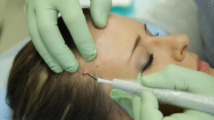
Papillomatous intradermal melanocyticNevus should be removed completely. Otherwise, it will begin to degenerate into a malignant tumor. Laser surgery is performed under local anesthesia and lasts only 5 minutes. After its implementation, you will go home on the same day, and the recovery period will be reduced by several times.
For work specialist uses a speciala device capable of producing laser radiation. He gradually cuts off the thin lamina of the neoplasm until it is completely removed. The depth and intensity of the radiation are regulated in each individual case. It all depends on the size of education, its features.
Preventive measures
Intradermal pigment nevus isquite unpleasant disease that can develop into a malignant tumor. However, if you follow some preventive measures, then such an outcome of events can be avoided. So, do not forget about these rules:
- Do not abuse visits to the solarium or stay there for too long.
- In summer, you are not under the influence of direct sunlight for a long time, especially in the period between 11 and 16 hours.
- Do not go to saunas or baths too often.
- You should be especially careful with the sun for those people who have fair skin, as it is more prone to the appearance of moles and melanomas.
- You can not injure a tumor.
If you find a mole that has drastically changed color, shape, or size, contact your doctor immediately. Be healthy!