


Череп человека – это костный каркас, который it has twenty-three bones in its composition. They perform the function of protecting the brain from various injuries. The skull is a component of the bone system and the musculoskeletal system. It consists of the brain and facial departments that perform certain functions. Each department has an external and internal base.
Оно образовано с помощью глазничных и носовой parts of the frontal bone, small and large wings, latticed bone and plates, temporal pyramids and the body of the main bones, lateral parts and lower portions of the occipital scales.

The base of the skull, the photo of which you see, has such a structure that some of its bones are partially joined by sutures or layers of cartilage tissue. They are called synchondrosis.
The external base of the skull is divided into sections andhas various protrusions and holes through which the nerves and blood vessels pass. The posterior part is the location of the external occipital protrusion. From it comes down the crest of the neck. Before the scales is a large opening of the occiput. On the sides it is limited to the occipital bone, and in front - wedge-shaped. The sublingual nerve has a condylar canal passing under the processes of the occiput, behind which is located the fossa, which passes into a non-permanent canal.

Not far from the large occipital openingThe base of the skull, closer to the front, is the pharyngeal tuber, and in the mastoid process there is an opening with the corresponding name, which is the exit point of the facial nerve, and the styloid process.
Нижняя поверхность каменистой части имеет яремную a hole and a hole with the same name. Through it, the cranial nerves pass. An external jugular vein emerges from this opening of the skull base. In front of him is a sleepy canal with an external hole, and a torn canal is located next to the tip of the rocky part.
The pterygoid canal passes just before the rootpterygoid processes and opens into the fossa. The oval and spinous apertures are on the sphenoid bone. Holes of the nasal cavity are called choana. They are located in front of the torn. Between the outer plate of the pterygoid process and the lower part of the surface of the large wing located on the sphenoid bone, there is a fossa fossa.
The posterior sections of the bone skull have the same holes, which lead to the canals. The incisors have cells behind which there is a incisive hole.
Череп – это полость, образованная плотно connected by bones, in which are located important for human body organs: the brain of the head, the initial parts of the respiratory and digestive systems and sense organs. In the skull, the vault, or roof, and the base, which is external and internal, are distinguished. The external base of the skull is formed with the participation of its lower surfaces - the brain and facial sections, which are subdivided into anterior, posterior and middle.
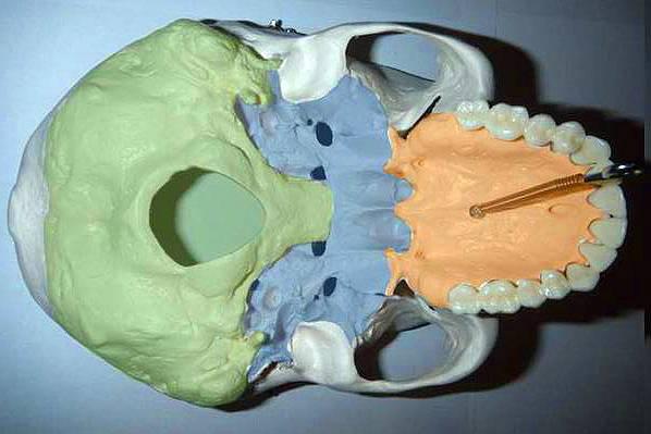
The anterior part originates from incisors andcaptures the posterior edge of the palatine bones formed by horizontal plates, which in front connect with the processes of the upper jaw, forming the bone skies. In its space, the incisal fossa is formed, from which the incisal canal begins. It leads to the lower courses of the nose. The structure of the base of the skull is such that a seam passes through the middle of the bone skull, and the palatine orifices: small and large - lead into the canal.
The middle section is occupied by the space between the sky anda large occipital foramen, and anterior margin. The lateral borders pass through the external auditory canal up to the process of the mastoid form. The outer base of the skull has two openings that open into the nasal cavity.
The posterior part is located between the anterior edge of the occipital large opening and the outer occipital mound.
В его состав входят парные и непарные кости.The first prevail. They are represented by the upper jaw, nasal, zygomatic, tear and palatine bones, lower shell of the nose. The second - a latticed bone, a vomer, a hyoid bone, a lower jaw. The skull base bones, of which the facial section consists, exert a tremendous influence on all sensory organs, the respiratory and digestive systems.

Being a solid skull allows an area,filled with air. They have unpaired bones. In addition, air is involved in providing thermal insulation. Such cavities are in the wedge-shaped, frontal, latticed, temporal bones and upper jaw.
A special role in the human body is allocateda sublingual arcuate bone located between the larynx and the lower part of the jaw and connected to the cranial bones by ligaments and muscles. With its help, the formation of the body and pair horns, from which go the styloid processes.
Верхние кости основания черепа плоские и are plates filled with bone substance. In his cells are the brain and blood vessels through which blood flows. Gyrations and grooves of the brain are formed due to unevenness of some bones of the skull.
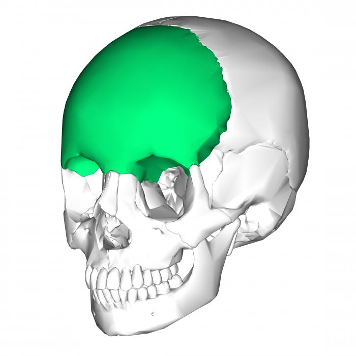
Он оберегает головной мозг от повреждений и is its protective frame. The brain of the skull is above the facial and has the shape of an ellipse. Its volume is 1500 cm. It consists of paired, parietal and temporal, bones, and unpaired - occipital, wedge-shaped and frontal. The latter consists of two scales, a bow. It is airborne. Here, the forehead and frontal hillocks form, due to which the walls of the eye sockets, the nasal cavity, the pits on the temples and in the anterior part are formed. With the help of the parietal bone, arches are formed, and with the help of the occipital bone - the base of the skull, the photo of which is presented to your attention.

Paired bone is a complex pneumaticthe temporal part. It carries out the formation of the calvaria, it contains organs of hearing. This bone forms a pyramid with a tympanic cavity and an inner ear.
It is located where the base of the skull, inits very center. The sphenoid bone has a body with processes from it, with a corresponding name, with large and small wings. The body has six surfaces that perform certain functions. This is the front, back, top, bottom and two side.
У основания большого крыла имеются отверстия round, oval and spinous. There are four surfaces of the wing, which are called the temporal, maxillary, orbital and brain. They are located arterial grooves and depressions. The medial side of the small wing has an inclined process. The space between the large and small wings occupies the upper orbital fissure.
It consists of the basilar, lateral parts andscales. When they are connected, a large hole is formed, which is called the occipital. The lower surface of the lateral part is provided with a condyle, above which the sublingual canal is located. Behind it is a fossa with a condyl channel at the bottom.
The center of the outer surface of the scales has an occipital protrusion. From it goes down the crest of the same name.
The outer base of the skull occupies most ofvault and has a frontal bone, which consists of the nasal, orbital part and the frontal scales. The nasal part of the front and sides of the lattice is limited to a lattice, which separates the right and left orbits. The anterior part of the frontal part in the middle has a line that passes into the nasal spine. On both sides of it (horizontal) is the aperture of the sinus of the frontal part of the skull.
Being a complex bone organ, the skull performs the following functions:
The newborn child has flat jaw bones,they are a collection of a huge number of bone beams without a clearly defined organization. Between them is loose connective tissue. Compact bone in peripheral zones is absent, it is replaced by the periosteum, represented by a thick layer.
Over time, the merging of the beams.Forms a continuous compact plate: first on the sides, then in the frontal and distal parts of the jaw. The dimensions of the facial bones are increasing. Of great importance in the growth of the facial bones is the base of the skull. The anatomy of their structure is such that the anterior cranial fossa is lengthened by the sutures, which separate the frontal and ethmoid, the latter and the main bone.
Ends growth at 10-11 years of human life.Subsequently, the frontal bone is pneumatised, and bone formation occurs on the outer surface. In girls, this occurs before the age of 13, and in boys, it reaches 14. With the growth of the base of the skull, the angle between the cranial fossae: the posterior and anterior decreases. This explains the fact that the vertical size of a person's face prevails over horizontal.
All the bones of the skull begin to develop withmembranous stage, followed by cartilaginous and final bone. The facial bones in their development bypass the middle stage. A feature of the structure of the skull of a newborn child is the presence of remnants of the membranous skull - the fontanelles, which are anterior, posterior and lateral.
On the front fontanel (largest) canobserve the respiratory movements, intracranial pressure (if it increases, the spring swells), dehydration of the baby’s body (if the disease falls, the spring falls).

Rear fontanel is smaller and fast.overgrown. A full-term newborn child often lacks lateral fontanelles that are present in preterm babies. But by 2-3 years of life they are overgrown.
The second feature is that both the inner and outer surface of the base of the skull has cartilage layers that are located between the individual parts of the bones.
The third feature. In newborns, pneumatic sinuses, processes, tubercles, jaws are not developed, there are no teeth.
The formation of sutures of the skull occurs by 3-5 years of human life. In general, he finishes growing in 25-30 years.
The skull is distinguished by gender, butIt does not matter. Age-related changes can spread to the entire base of the skull. The anatomy of its structure is such that the crests and the bone substance of the spongy structure begin to dissolve, the cranial bones become light and fragile. The shape of the skull may change under the influence of mechanical factors.


