В организме человек огромное количество костей.Especially a lot of them in such mobile areas of the body, as the foot and wrist. Dozens of bones connected by tendons help to perform work that is inaccessible to animals, except perhaps for monkeys. The complex system of hands and feet, although it has a large amount of connective tissue, is subject to various injuries and diseases. The most common is a fracture. The concept is related to the fracture of the bone and the possible displacement. In the hands and feet, as already mentioned, a large number of these organs, which do not differ in size, therefore, their treatment takes a lot of time. Scaphoid most prone to disease and injury.
Leg bones are presented in huge quantities.Some of them are united by the common name of the foot. Scaphoid bone belongs to this group. It is located between the ram, cuboid and intermediate sphenoid bones. It is this place of the foot, excluding the toes, that is most often prone to fractures.
The bones of the foot, the anatomy of which is represented by threedepartments, quite numerous: tarsus, metatarsus and fingers. The metatarsal bones of the foot contain scaphoid in their rows. It is located near the inside of the foot. At its edge is located tuberosity of the navicular bone, directed downwards. In medicine, this feature is used to determine the arch that the foot has. X-ray helps to understand the composition of this part of the body.

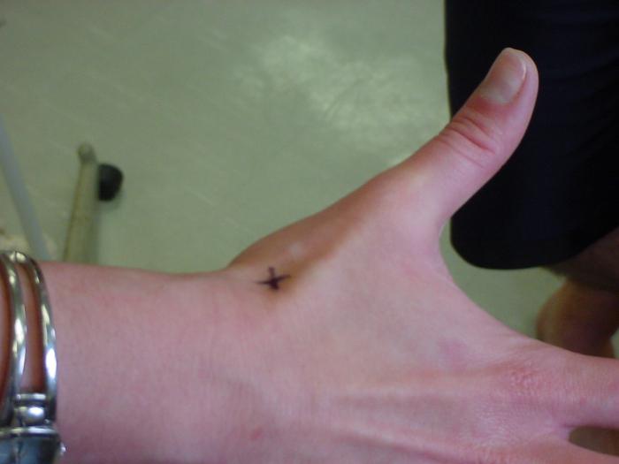
Scaphoid is also located incyst. It refers to the small bones of the wrist. It is she who is predisposed most of all to the fracture, since she is on the edge of the palm. It is interesting that the person who broke this bone does not feel any particular pain and can only feel bruise, even if it is strong. Therefore, it is quite dangerous. If you do not see a doctor, then there can be serious consequences. For example, the navicular bone may incorrectly fuse.
The wrist consists of 8 bones.They form 2 rows, in each of which there are 4 of them, located between the metacarpal bones and the forearm. Scaphoid is easy to grope because of its location. It is located between the tendons of the long extensor of the big toes and the long abductor.
In addition to fracture, the navicular bone of the foot is susceptible.and other injuries and illnesses. For example, Kellerra disease. Osteochondropathy can be a herald of this disease. It affects all the bones of the foot. Gradually destroys tissue. During the illness, a small amount of blood flows to the bones, which means there is not enough oxygen and nutrients. Consequently, cells that did not receive a sufficient amount of this gas and other necessary components gradually die off. However, this happens in the case of Keller's disease, without the intervention of the infection.
Keller's disease can not occur by itself.For her, there are several reasons that somehow interfere with the passage of blood to the bones. Most often it is a foot injury, for example, severe injury or fracture. Also those who wear uncomfortable small shoes are susceptible to this disease. Osteoarthritis and arthritis are diseases that also lead to Keller's disease. In addition to the above reasons, congenital deformities of the foot bones can also lead to deterioration. Flatfoot - one of the main defects. But the reasons that directly affect the appearance of the disease have not been found even today.

Leg bones are susceptible to two types of Keller disease. It all depends on which part of the foot does not receive enough nutrients and oxygen.
With the defeat of the navicular bone diseaseKeller's disease is called 1. If blood does not flow to the heads of the third and second metatarsal bones, which leads to their change, then this is called Keller's disease 2.
In addition, several stages are distinguished:
При первой стадии погибают костные балки, которые and perform the role of structural elements of the bone. Next comes the formation of new parts of the bone tissue, which often break due to poor strength. Then the bone beams dissolve. And the last stage is fully consistent with the name.
It is necessary to treat the bone of the foot.Their anatomy is extremely difficult, so they are not easy to cure. With Keller's disease 1, a fracture of the navicular bone most often occurs. It can be taken for injury, and the disease is extremely difficult to detect. Unless, by chance, the ill will go to a doctor. After there is a course of treatment. The bone with the same name is also in the hand, but this will be called Ireiser’s disease, although the treatment principle will be the same.
Conservative therapy is one of the mosteffective treatment methods. A plaster cast is also applied. It is not recommended to move the foot itself, as it is difficult to fix such a small and non-standard bone. After removing the plaster, in order to maintain the result, you need some time to walk on crutches or with a cane, special insoles are sewn to the children. Drugs can accelerate healing. Thermal procedures are very helpful.

Нельзя после снятия гипса заниматься спором.The foot needs constant rest. There is also the possibility of improper accretion and the formation of a false joint that is difficult to cure. It will take an operation. Therefore, the rehabilitation process must be treated with the utmost attention. In addition, you can take only those drugs that are prescribed by the doctor, otherwise you can only make the leg worse. We can not neglect the advice of a doctor, because each person has his own characteristics of the organism. Some bones are fragile from birth, so they should pay special attention to the treatment of this disease.
As already mentioned, the navicular bone of the arm andfoot more than others at risk of fracture. This is due to the fact that both on the foot and on the hand, the bone is located in such places with which injuries occur most often. If we look at the statistics, in the case of a wrist fracture, 61–88% suffer from the scaphoid.
But why is this bone fractured?As practice shows, many are injured, falling on his hands. In this case, the load almost completely falls on the bone. The fractures themselves also differ: intra-articular and extra-articular.

Scaphoid very often injured.But after the fracture, it almost does not hurt. Most simply do not notice the inconvenience, thinking it is just a bruise. However, it is necessary to consult a doctor as soon as possible. Scaphoid is poorly treatable, and if you do not have time to its accretion, but there may be irreparable consequences. Unfortunately, not all go to the hospital. Most often, a fracture is detected randomly. There are some symptoms that can help recognize the injury:
As with a bone fracture on the wrist,Injury of the navicular bone on the foot suffers greatly from the foot. X-ray helps to detect the cause of pain. Initially, a 3D projection is performed on the device, for which purpose zones are explored in three projections. At the final stage, a fracture (fracture) of the navicular bone is clearly visible. All this is due to the fact that the navicular bone is extremely difficult to treat, it is surrounded by other organs. To correctly and accurately put a plaster cast, just need a 3D projection.

Have their own subtleties.For example, fingers should be clenched into a fist. If the fracture is not immediately visible using X-rays, but by all indications it is, then the victim wears a plaster cast for about 2 weeks, then his brush is re-checked. The thing is that during this period resorption occurs and the crack will be clearly visible, if, of course, it is present at all. Actions help establish the diagnosis and prescribe treatment.
Scaphoid bone on the wrist is often the casebroken, which is extremely difficult to detect. To detect a fracture, one has to resort to 3D projection. But the treatment of a fracture is much longer and cumbersome. Bone consolidation is purely due to endosteal corn, which is formed extremely slowly, and requires a large amount of nutrients (blood). Possible displacement of the distal fragment. All of the above leads to the formation of a false joint, and thus complicates the already difficult treatment.
The easiest way to build a navicular bonehands - this is the imposition of a plaster cast. The most common, it is used in 90–95% of cases. The overlap occurs from the heads of the metacarpal bones down to the elbow joint, with the capture under the bandage of the phalanx of the little finger is considered mandatory. The brush remains stationary, but for the convenience of the victim, its position has the form of a slight extension. Immobolization of the brush lasts about 11 weeks. If the fracture occurred with a tubercle, then it is only 4 weeks. After removing the plaster cast, an x-ray is mandatory, which will show whether the accretion has occurred correctly. If a slit is detected, the plaster cast is applied again, but for 1–2 months, with the control of accretion occurs every month. After the end of treatment, a course of recovery occurs.
The disadvantages of conservative treatment include:
If the fracture is found only byafter 3 months, it is considered old. By this time, the false joint has time to grow. This complicates the treatment. With the help of X-rays find the place of the fracture, as well as determine the presence of cystic cavities and diastase between the debris. In this case, the imposition of a plaster cast cannot help. One of the most famous methods is used:

The method was invented back in 1928.It is used with non-accrued fractures and false joints of the navicular bone. Rear-beam access is used for anesthesia during surgery. Without damage, without touching the radial nerve, access to the wrist joint occurs. Dissecting its capsule helps to detect the false joint. After the operation is completed, a plaster cast is applied in the same way as described above. It is necessary to take about 14 days with it. Then, remove the stitches and use a circular dressing. The role of the bone plate is often performed by a spongy graft.
One of the most efficient operations.But it is quite simple. For it, the blood field is exsanguinated, however, thus the blood supply practically does not deteriorate. Stabilize the navicular bone with the help of the spokes. A graft is wedged into the bone. Pre-placement of the spokes does not allow debris to mix. Immobolization takes about 10 weeks. The removal of the needles occurs only after 8 weeks.
Как уже было сказано, кости предплюсны наиболее prone to various kinds of injuries. Most often a fracture occurs after the fall of a heavy object on the foot. Sometimes not one bone suffers, but several, since they are located in close proximity to each other and have small sizes. As with the navicular bone of the wrist, do not delay treatment. However, the foot is much easier to treat. A fracture of the navicular bone occurs directly, either due to the fall of an object with a large weight, or due to squeezing between others. The foot bones are quite diverse, their anatomy numbers dozens of species.

Reveal a fracture of the navicular bone of the foot mucheasier than a brush. With an injury of this kind it is almost impossible to move normally, there is constant pain. In addition, the circular motion of the foot reveals a final fracture, the bone makes itself felt. But almost always, the injury of the navicular bone is combined with the injuries of other bones of the foot and, in particular, the tarsus.
To know the size and location of the crack,it is enough to make an x-ray in 2 projections, and not in 3, as was the case with the navicular bone of the hand. If there is no displacement, then the usual plaster cast is applied. But if it happened, reposition is performed. In the most difficult cases, open reduction is performed. A plaster cast is applied an average of 4 weeks.
In conclusion, we can say that the scaphoidmore than other wrist and foot bones are prone to injury. For her treatment takes a long time, often have to resort to operations. However, bone fusion on the foot is much faster and easier. It is quite difficult to detect a fracture on a cyst, and most often it happens by chance. Scaphoid foot bone in the case of a crack sore.


