


The lower jaw of a person (lat.mandibula) is an unpaired mobile bone structure of the facial cranium. It has a well-defined central horizontal part - the body (mandibulae of Latin origin) and two outgrowths (branches, Latin ramus mandibulae) that extend at the edges of the body of the bone.

She takes part in the chewing processfood, speech articulation, forms the lower part of the face. Consider how the anatomical structure of the lower jaw is related to the functions performed by the bone.
During ontogeny, the structure of the lower jawa person changes not only in utero, but also postnatally - after birth. In a newborn, the body of the bone consists of two mirror halves connected in a semi-mobile manner in the center. This middle line is called the chin symphysis (Latin symphysis mentalis) and completely ossifies by the time the child reaches one year.
The halves of the lower jaw are curved arched,located convex to the outside. If you outline the perimeter, the lower boundary of the body - the base - smooth, and the upper has alveolar depressions, it is called the alveolar part. There are holes in it, where the roots of the teeth are located.
The branches of the jaw are located by wide bone plates at an angle of more than 90 ° C to the plane of the body of the bone. The place of transition of the body into the maxillary branch is called the angle of the mandible (on the lower edge).
From the side turned outward, the anatomical structure of the lower jaw is as follows:

Such features of the structure of the lower jaw, asthe size and morphology of the chin protrusion, the degree of curvature of the bone, form the lower part of the face oval. If the tubercles strongly protrude, this creates a characteristic relief of the chin with a dimple in the center.

In the photo: the lower jaw affects the shape of the face and the overall impression of it.
On the inner side, the relief of the mandibular bone (its body) is due mainly to the fixation of the muscles of the bottom of the oral cavity.
The following areas are distinguished on it:
The upper third of the jaw's body has thin walls, bounding the dental alveoli. The border is an alveolar arc, which has elevations in the places of the alveoli.
The number of cavities corresponds to the number of teethlower jaw in an adult, including the "wisdom teeth" appearing later, 8 on each side. The pits are septated, that is, separated from each other by thin-walled partitions. In the area of the alveolar arch, the bone forms protrusions corresponding to the dilatations of the dental holes.
The anatomy of the bone in the region of the branches is determined by the muscles fixed to them and the mobile joint that connects it to the temporal bones.
Снаружи, в районе нижнечелюстного угла, имеется an area with an uneven surface, the so-called chewing tuberosity (Latin tuberositas masseterica), on which the masticatory muscle is fixed. Parallel to it, on the inner surface of the branches, there is a smaller porphyritic pterygoid (Latin tuberositas pterygoidea) - the location of the pterygoid medial muscle.

On the central part of the inner surfacethe mandibular opening opens the opening of the lower jaw (Latin foramen mandibulae). The elevation - the mandibular tongue (Latin lingula mandibulae) partially protects it from the front and medially. The hole is connected by a channel passing through the thickness of the bone spongy substance with the chin opening on the outside of the mandibular body.
Above the pterygoid tuberosity is locatedelongated depression - mandibular hyoid furrow (Latin sulcus mylohyoideus). In a living person, neural bundles and vessels pass through it. This furrow can be transformed into a canal, then it is partially or completely covered by a bone plate.
On the front edge of the inner side of the branches, starting just below the level of the opening of the lower jaw, descends and continues on the body of the mandibular shaft (Latin torus mandibularis).
At the ends of the branches two processes are well pronounced:
Between the processes there is a deep notch - a notch (Latin incisura mandibulae).
Anatomy of the end sections of the branches of the lower jawprovides its good mobility and articulation with the bones of the facial skull. Movement is possible not only in the vertical plane, the jaw is also shifted back and forth and from side to side.

The temporomandibular joint forms, respectively, two bones: the temporal and lower jaw. The structure (anatomy) of this joint allows it to be attributed to the type of complex cylindrical joints.
The maxillary articular fossa of the temporal bone is in contact with the anterior-superior portion of the condylar process of the jaw. That it should be considered the true articular surface.
The cartilage meniscus inside the joint divides it into two"Tier". Above and below it there are not intercommunicating gaps. The main function of cartilaginous pads is to absorb when rubbing food with teeth.
The temporomandibular joint is strengthened by four ligaments:
The first one is the main one, the rest carry an auxiliary supporting function, since they do not directly cover the joint capsule.
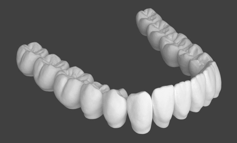
The anatomical structure of the teeth of the lower jawdetermined by the necessity of closing and contact with the upper row of teeth. Their specific location and interaction is called a bite, which can be:
Changes in occlusion adversely affect the process of chewing food, provoke speech defects, distort the facial contour.
В норме строение и рельеф поверхности the mandibular row of teeth ensure their close contact with the same maxillary teeth. Mandibular incisors and canines partially overlap with similar upper teeth. The outer tubercles on the chewing surface of the lower molars lie in the pits of the upper ones.
The lower jaw is not monolithic. The presence of channels in it, areas with different density of bone material causes typical injuries due to injuries.
Common sites of mandibular fractures are:
Since the bone is thickened in the area of the mental symphysis, and at the level of 2 and 3 pairs of molars it is strengthened by the internal ridge and external oblique line, the lower jaw is rarely broken in these places.

Another damage option that does not affect itself.the bone, and the temporomandibular joint, is dislocated. It can be provoked by a sudden movement to the side (from a blow, for example), excessive opening of the mouth, or attempts to get through something hard. The articular surfaces in this case are displaced, which prevents normal movements in the joint.
The jaw must be replaced by a trauma specialist,to prevent excessive stretching of the surrounding ligaments. The danger of this injury is that the dislocation can become habitual and recur with little effect on the jaw.
The mandibular joint experiences permanentloads throughout a person’s life. He is involved in eating, talking, important in facial expressions. His condition can affect lifestyle, diet, the presence of systemic diseases of the musculoskeletal system. Injury prevention and early diagnosis of articular problems is a prerequisite for the normal functioning of the lower jaw throughout a person’s life.


