


Skull X-ray is one of the available andinformative diagnostic methods. With it, you can check the state of the internal structures and bone elements. The value of the study is the ability to diagnose the patient's condition after a head injury, to detect the tumor process, the presence of pathological fluids.

Craniography allows the doctor to detect the following points:
X-rays of the head allow you to obtain diagnostic field data on film, on a monitor screen. If necessary, they are stored in the X-ray memory.
During the review X-ray estimatethe state of the brain as a whole. Sighting craniography allows you to make sure in the state of a certain part of the head, to clarify its functionality over time with several pictures taken in a row.
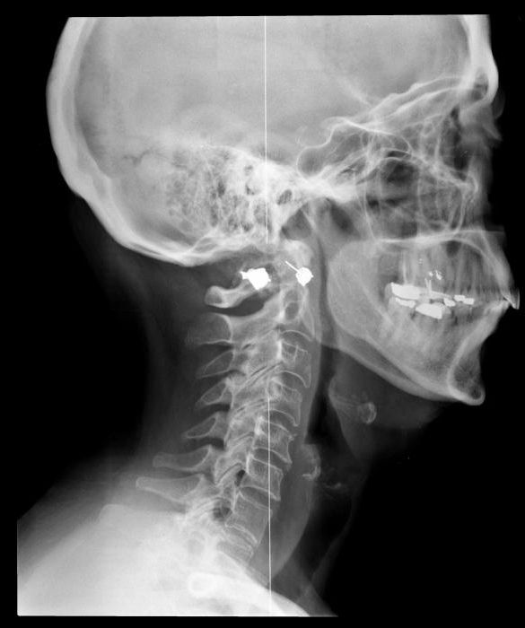
An aimed X-ray of the head is carried out in order to detect fractures in such bone elements:
Aim pictures allow you to see:
X-ray of the skull is done on the basis of complaintspatient or those changes in the patient's condition that were seen by the doctor during the examination. You need to be prepared if the specialist sends to craniography in case of complaints of trembling in the limbs, cephalgia, darkness or shading before the eyes, nosebleeds, pain during chewing, loss of vision or hearing.

Mechanical indications may also be indications.head injuries, asymmetry of facial bones, fainting, suspicion of malignant tumors, endocrine apparatus pathologies and congenital anomalies.
Pregnant women and women during lactation do not carry out an X-ray of the skull bones. The following specialists may send for the procedure:
Special training this survey method is notrequires There is no restriction (in drinking, food, medicine) before the procedure. Before the subject takes a place in the X-ray unit, he needs to remove metal things, dentures (if possible), and glasses. Further, depending on the study area, the patient lies down on the couch, sits down or stands.

A lead apron is worn on the subject tothe body below the head did not receive excess radiation. The head is fixed with the help of special fixers so that the area of the examination remains immobile for the entire period of diagnosis. Sometimes they use fasteners or bandages, sometimes ordinary sandbags.
If necessary, the radiologist can take not one, but several pictures. In addition, the position of the body can be changed to perform an X-ray of the skull in several projections.
Speed and claritythe images on them depend on the modernity of the radiological apparatus used. In exceptional cases, the answer may be issued to the subject immediately after the procedure, but in most cases it is required to wait until half an hour. In public medical institutions, the decoding of the results can take up to several days.
The decoding of the image contains data on the shape of the cranial bones, their condition, size, correctness of the anatomy, the contents of the paranasal sinuses, the state of the cranial sutures, the bones of the pyramid of the nose.

X-ray skull in 2 projections that shows?For more informative results, the radiologist conducts a study in several projections (usually in the anterior and lateral). This allows you to more accurately determine the size of pathological structures, their localization, the state of the bones, the presence of displacement.
X-ray of the skull is accompanied by a lowirradiation of the patient’s body (approximately 0.12 mSv). This indicator is less than 5% of the dose that is allowed to be received by a person per year. For comparison, we can say that a person receives the same amount of radiation while relaxing under the sun on the beach in one hour.
However, an x-ray of the head (as shown by this method, described above) is not recommended more than 7 times a year.
X-ray diagnosis is carried outsolely by indication and its purpose is to determine the presence of a deadly disease. That is why there are cases of greater patient radiation than indicated in the medical literature. For example, a skull fracture is considered an emergency. If he is suspected, diagnostics is performed even during pregnancy. Women carefully cover their breasts and abdomen with lead apron.
Child's X-ray Skull - a procedure requiringmore thorough approach. In most cases, the specialist prefers ultrasound. X-ray diagnostics is used with extreme measures, since the bone elements of the brain are still in the stage of their growth and formation, and excessive exposure can lead to negative consequences.

Frequent indications are head injuries, includingincluding a generic, and a fracture of the skull. The procedure is similar to the examination of adults. The only problem is the need to be in the same position during manipulation, which is very difficult for children. It may require the presence of parents or taking sedative, hypnotic drugs before diagnosis.
One of the indications for craniography. Injuries can have a scalped, ragged, cut, chopped, blunt character depending on the way they occur. The main reasons are:
If there is damage only to the soft tissue, this condition is called a head contusion. In violation of the functionality of the internal structures talk about traumatic brain injury.
Пострадавший чувствует боль в месте травмы и there are no other manifestations - this state does not require the help of doctors. Apply to the place of damage cold. If there is bleeding, nausea and vomiting, pain in the neck, dizziness, hospitalization and the help of specialists is necessary.
An emergency requiring urgent assistance and calling the medical team to the place of injury may be accompanied by the following manifestations:
Awareness of what to do whenhead injury, can save the life of not only someone from outsiders, but also relatives, relatives. First of all, it is necessary to provide the victim with rest before the ambulance team arrives. The person should be laid on a bed with a slightly raised head end, if possible in a dark room. Must be near someone.

If vomiting is present, do not allow to get uppatient, and turn his head on its side and substitute a container for vomitus. In the case of convulsive seizures, a person is turned on his side with his whole body, a solid, but not a metal object is inserted between his teeth so that the tongue does not fall.
Bandage should be applied to the wound.by hand if there is bleeding. If you suspect a fracture to put pressure on the skull is not necessary. In parallel, you need to monitor the presence of the pulse and respiration. If there are no signs of life, start cardiopulmonary resuscitation.
Никакие медикаменты, даже обезболивающие, давать the victim does not need an ambulance before the arrival, as this may hide the true picture of the condition. It is necessary to clarify the state of memory of a person by asking him a few questions about his name, relatives, the place where he is at the moment. Apply cold to the bruise.
Even having a good knowledge of the ability to providefirst aid, you need to be calm and reasonable to leave panic aside and soberly assess the situation. And the best option, if possible, is to prevent injury than to restore the victim’s health.


