


The structure and function of different parts of the body, includingincluding bone compounds, studies anatomy. The elbow joint belongs to the bone joints of the free upper limb and is formed as a result of the articulation of individual parts of 3 bones: the humeral, ulnar and radial.
The elbow joint is an unusual bone joint that unites the shoulder and forearm.

A complex joint is one in which more than two articular surfaces take part. There are three elbows:
Combined joint refers to those compounds in which several independent joints are joined by one joint capsule. In the elbow in one capsule three independent are combined.
The anatomy of the elbow joint of a person is very unusual; it combines 3 different types of joints in one joint:
The structure of the joint allows you to perform a specific set of movements. This is flexion, extension, rotation (pronation and supination).
The joint capsule surrounds 3 joints. It is fixed in front and sides.
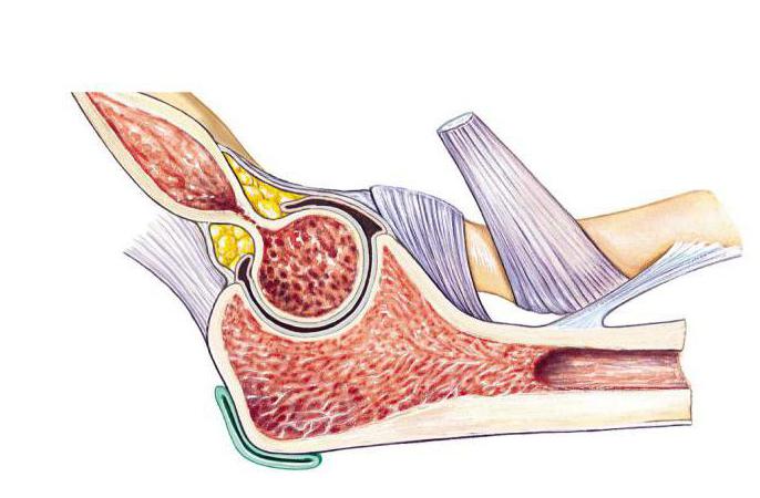
Each bone compound is a complex and thoughtful anatomy. The elbow joint is strengthened by ligaments, which provide its protection and movement in different planes.
The ulnar collateral ligament starts from the base of the humerus (medial condyle), ends at the ulna (block-like incision).

The annular and square ligaments fix the radius and ulna.
Lumpy tendons of the elbow joint are attached by lumps. The anatomy of this compound is called the “ulnar bone head”. It is she who most often suffers from injuries and injuries.
Besides the main ligaments of the joint, in functionbone fixation is involved and the interosseous membrane of the forearm. It is formed by strong beams that connect the radius and ulna. One of these beams goes in the opposite direction from the others, called the oblique chord. It has holes through which vessels and nerves pass. Oblique chord is the beginning for a number of muscles of the forearm.
There are some unusual bone in the human body.compounds. Anatomy studies them all. The elbow joint is unusual in its own way. It is protected by a good muscular frame. The harmonious work of all muscles ensures the smooth operation of this bone junction.
All the muscles affecting the elbow joint can be divided into 3 groups: extensors, flexors, rotators (perform pronation and supination).
The extensors of the joint are the triceps muscle of the shoulder (triceps), the fascia tensioner of the forearm, and the ulna.

Pronators - brachiocephalus muscle, round pronator, square pronator make rotational movements in and out.
The instep supports - the biceps muscle of the shoulder, the instep support, and the shoulder-shoulder muscle rotate the forearm from the inside.
Performing physical exercises that strengthen the listed muscles, it is important to remember about safety. The elbow joint is very often injured in athletes.
It is very important for the joint to get in timenutrients that come to him with blood. It comes to all joints and muscles from a group of arteries. They consist of 8 branches, which are located on top of the articular capsule.
The network of arteries supplying blood to the joint consists of vessels called "anastomosis."
Topographic anatomy of the elbow joint is a very complex diagram of the connection of vessels. Thanks to this scheme, the blood supply to the joint is uninterrupted. Outflow is carried out through the veins.
What makes the process of movement in the joint possible? There are special nerve structures that carry out the innervation of the muscles. This is the radial and middle nerves. They run along the front of the elbow.
The elbow joint is very vulnerable, as it is constantly subjected to physical exertion.
Very often, to understand the cause of the pain, the doctor prescribes additional research. This may be radiography, MRI, ultrasound, tomography, arthroscopy, elbow puncture.

The main method for diagnosing diseases of the elbow is radiography. The pictures are taken in two projections. They allow you to see all the changes in the bones.
To identify diseases of the soft components of the elbow, doctors use other methods of research.
Regular pains in the elbow area may indicate that there are any violations. After the examination, the most frequent diagnosis is arthrosis. It happens and arthritis, and much more.
It is much less common than in the knee orhip joints. The risk group includes people whose work is associated with increased loads on the elbow joint, those who have suffered an injury or surgery on the elbow, endocrine or metabolic disorders, and arthritis.
Main symptoms: constant aching pain that occurs after physical activity. It takes place after a rest. Clicking or crunching in the elbow. Limit the amplitude of motion.
Inflammation of the joint. The possible reasons are many. They can be infections, allergic reactions, high loads on the joint, malnutrition.
The form of arthritis can be acute or chronic.
Main symptoms: persistent pain, skin flushing, swelling, limitation of joint mobility.
Most often the elbow joint affects rheumatoidarthritis. Its symptoms are: stiffness in the morning, symmetrical arthritis (both joints are inflamed), chronic nature of pain, involvement of smaller joints (hands, ankles, wrist, knee) into the painful process.
Frequent disease in people whose activity is associated with high loads on the elbow (tennis, golf, wrestling).
There are 2 types: lateral, medial.

Main symptoms:pain in the area of an injured epicondyle that spreads to the muscles of the forearm (anterior or posterior). At the onset of the disease, pain occurs after exercise. In the future, the pain is felt even by minimal movements.
Inflammation of the articular bag. Most often occurs in people whose activity is associated with permanent injuries of the back surface of the elbow.

Unwanted physical impact on the elbow can cause injury. These include dislocation, bone fractures, sprain, joint hemorrhage (hemarthrosis), muscle damage, rupture of the articular capsule.
Listed injuries and illnesses most oftenmeet in everyday life. In order to protect yourself from them, you should take preventive measures: avoid excessive loads, give yourself time to rest, prevention of traumatic situations at work is important, maintaining a diet, you need moderate physical training and articular gymnastics.


