


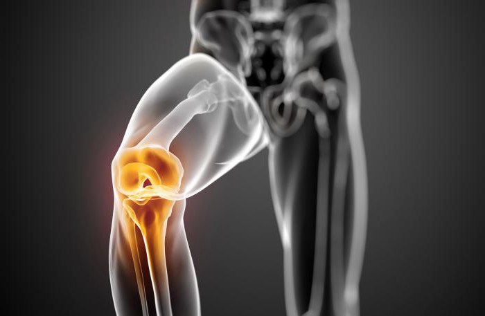
In the human body there are 206 bones, a largesome of which do not exceed several cubic centimeters in size. The most painful and massive bone in the body is the femoral. Its structure allows us to walk straight and not fall. Through the knee joint, the femur connects with the tibial and peroneal, forming a free lower limb.

Anatomy of human lower limbs includesyourself bones, muscles, ligaments, joints and fascia. This is if you understand seriously and thoroughly. But for this article there will be enough a small excursion into the structure of the leg. So, a person's lower limb is divided into the thigh, shin and foot.
The basis of the thigh is the femur bone.It is overlapped in layers by muscles, thanks to which a person can walk, stand, run, swim and much more. Working on the principle of a lever, they act on the hip or knee joint. The anatomy of the myofibrill allows them to stretch and shrink, adjusting to the needs of the body.
The core of the tibia - tibial and peronealbones. Between themselves, they are connected by a joint and connective tissue membrane in which the vessels pass. On top of this design is covered with several layers of muscles, which continue on the foot.
The ankle and the foot are parts of the body,who are under constant stress. A relatively small area of the sole holds the weight of the whole body (and sometimes it can reach three hundred kilograms). The foot consists of the calcaneus, tarsus and metatarsus, which are covered with fasciae and muscles. Also this area is abundantly supplied with blood so that the muscles always have a supply of oxygen.
Что же все-таки представляет собой анатомия human knee joint? For a first year student at a medical university, this is one of the most difficult questions, because you need to remember all the structures that make up this joint:
- bones (as a basis);
- muscles (contracting, they change the position of the leg);
- nerves and blood vessels (feed tissue and transmit information from the brain to the periphery);
- menisci (form the surface of the joint);
- ligaments (hold the bones together);
All of the above components of a healthy person work harmoniously, as a single mechanism. But it is necessary to "break" at least one component, and a smooth gait will not work.

The large bones of the knee are the femoral andtibial But besides them there is still a small rounded bone, located separately from the rest. It is called the patella or patella. On the diaphysis of the femur are spherical elevations - condoms, covered with cartilage for better sliding. They are the upper part of the knee joint. The lower part is formed by a flat head of the tibia, also covered with cartilage tissue.
The fibula is not long enough toshape the knee joint. The anatomy of its head allows it to adhere to the tibia in such a way that the shin can be rotated slightly without fractures. The thickness of the cartilage covering the articular surface reaches five millimeters. It is necessary to reduce the friction force, as well as depreciation.

As mentioned above, in addition to bones and musclesthere are still ligaments of the knee joint. Their anatomy is very entertaining, since it is these strips of tissue that keep all parts of the mechanism together. To strengthen the joint capsule, there are medial and lateral collateral (enveloping) ligaments on the sides of the bones. Between the upper and lower articular surfaces are cruciate ligaments. Topographically, one can distinguish the anterior and posterior ligaments, limiting excessive bending and extension of the knee.
Ligaments are important elements of the joint. They stabilize it, make the gait more firm and allow to avoid dislocations.

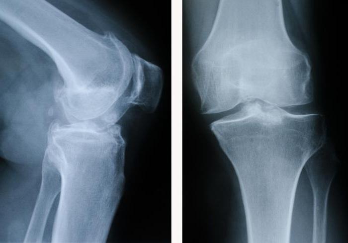
Если вы посмотрите на снимок коленного сустава, then besides the bones you will see two small formations. These are dense connective tissue formations - menisci. They are located between the femoral and tibial bone.
The two main functions of the meniscus are:
- increase the surface area of the joint for a better distribution of human weight;
- improving the stability of the knee joint, along with ligaments.
Для того чтобы представить какую роль выполняю menisci, you need to imagine the ball, located on a smooth flat surface. If there is nothing between a ball and a plateau, it will roll. Nature does not tolerate emptiness, and therefore the inside of the joint should not be empty either. Connective tissue fills the space between the articular surfaces, increasing their area and protecting from excessive loads. Damage to the meniscus is fraught with joint inflammation and destruction of cartilage
From the front of the thigh to the knee jointdown muscles extensors. One end is fixed to the femur or pelvis, and the other goes into the tendons and weaves into the joint capsule. The main one in this muscle group is the quadriceps. When it contracts, the leg unbends in the joint.
По задней поверхности бедра расположены flexor muscles. They also begin on the belt of the lower extremities, and end in the joint capsule in the form of tendons. When this group cuts, the leg bends.
Nerve fibers, arteries and veins braid like a networkknee-joint. The anatomy of the vessels in this area is not fundamentally different from the rest of the body. The artery, accompanied by two veins, passes along the back surface of the joint, supplying the lower leg and foot.
Next to them is the popliteal nerve,which is a continuation of the sciatic nerve. Slightly above the knee joint, it is divided into two parts and already in this form it descends to the lower leg and foot. Thanks to him, the free lower extremity is obtained by sensory and motor innervation.
Когда случается травма колена, травматологу нужно using physical and hardware methods to find out what is damaged and how serious it is. It is not enough to just look at the knee joint.
1. Lachman test or drawer symptom.Conducted to determine damage to the anterior cruciate ligament, if a picture of the knee joint could not be done. To do this, the patient is placed on his back and bend the injured leg at the knee joint thirty degrees. Then the doctor fixes the thigh and simultaneously moves the lower leg forward. If movement is possible, then the ligament is damaged.
2. Бесконтактный тест.If for some reason the doctor cannot touch the patient (for example, there is an obstacle in the form of obstruction or water between them), and the examination is necessary, then this technique can determine the presence of a complex injury. For this, the patient, lying on his back, with both hands, holds on the hip of the injured leg near the knee joint. Then the victim tries to raise the lower leg without extending the knee. If he succeeds and at the same time the tibia does not move, then the ligament damage is.
3. Test rear sag.In order to detect damage to the posterior cruciate ligament, you can also not do an x-ray of the knee joint. The technology of this study is simple, reliable and widely available. The patient should be asked to lie on his back and bend his knees at an angle of ninety degrees. If at the same time the tibial bone moves backwards, then the ligament is damaged.
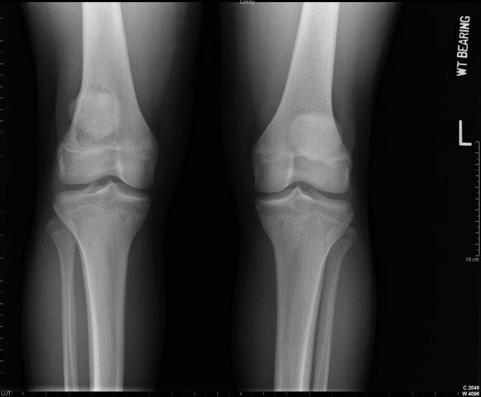
The most common survey methodbones is radiography. If the patient complains of joint pain after a fall, fever, swelling and hematoma, it is advisable to check if there is a fracture. An x-ray of the knee allows you to see the bones, soft tissues and tendons. Looking at the picture, a traumatologist can make a diagnosis: fracture, dislocation, sprain, damage to the patella, arthrosis, arthritis, a tumor or cyst, osteoporosis or osteomyelitis. These are the most common diseases affecting the knee joint. Photo, of course, can be of different quality, rigidity and size, but for a specialist it is not difficult.
In order to eliminate rheumatoid arthritis,degenerative pathology and joint injury, ultrasound can be performed. Another positive thing is that the patient does not need to undergo preliminary training (hunger, drink plenty, etc.) before examining the knee joint. Its anatomy allows you to look inside the articular bag, see the menisci, the surface covered with cartilage, bone formation.
Ultrasound allows you to view the knee from all sides. For a clear picture you need to properly lay the patient:
- on the back with straightened legs (the front and side walls of the joint are clearly visible);
- legs bent at the knee joints (menisci are visualized);
- in position on the abdomen (for examination of the posterior wall of the joint).
This procedure can be performed in almost any medical institution.


